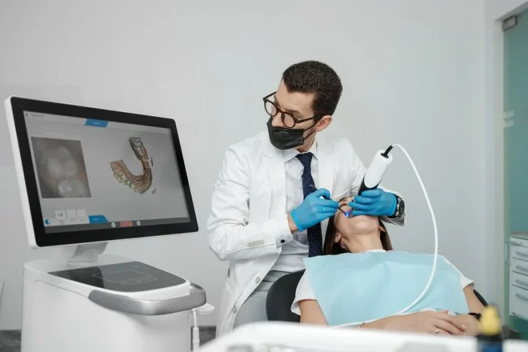
Biostimulation vs. Hyaluronic Acid: How Practitioners Select Dermal Fillers for Long-Term Tissue Restructuring
 Johnathan Parker |
August 26, 2026 |
3:53 pm
Johnathan Parker |
August 26, 2026 |
3:53 pm
In aesthetic medicine, the paradigm of facial volume restoration has undergone a fundamental shift. For years, the default approach to soft tissue augmentation relied almost exclusively on space-occupying gel matrices designed to immediately replace lost volume. Today, clinical focus has moved toward long-term tissue restructuring, prompting a clear distinction between immediate, temporary volumetric filling and biostimulatory neocollagenesis.
Selecting between hyaluronic acid (HA) gels and biostimulatory agents requires an understanding of distinct biochemical mechanisms, degradation kinetics, and structural outcomes. While hyaluronic acid remains the dominant option for immediate hydration and predictable, reversible volume replacement, biostimulators trigger a targeted, low-grade inflammatory cascade that encourages native type I and type III collagen synthesis over several months.
Mechanisms of Action: Direct Filling versus Endogenous Neocollagenesis
Hyaluronic acid dermal fillers function primarily through physical hydrogel retention and water binding. HA molecules contain repeated disaccharide units that bind up to 1,000 times their weight in water, creating an immediate, cohesive cushion within the targeted tissue plane. Rheological properties—specifically elastic modulus ($G’$) and viscosity ($tan delta$)—dictate how a specific HA product behaves under shear stress and compression. Higher $G’$ fillers resist deformation, making them ideal for supraperiosteal placement along the zygomatic arch or mandibular angle. Conversely, low $G’$ formulations integrate smoothly into dynamic areas without clumping.
Biostimulators operate on a fundamentally different physiological pathway. Instead of acting purely as an inert space-occupying gel, microparticles suspended within a carrier medium induce a controlled response upon injection. Macrophages absorb the carrier vehicle—typically a carboxymethylcellulose (CMC) gel—while host fibroblasts surround the exposed microparticles. Why does this matter? This cellular interaction prompts fibroblasts to deposit new extracellular matrix (ECM) components, predominantly type I collagen, along with elastic fibers.
Research published by the American Society for Dermatologic Surgery (ASDS) emphasizes that this endogenous restructuring continues long after the initial carrier vehicle has been reabsorbed by the lymphatic system. Consequently, while hyaluronic acid volume correlates directly with the volume of product injected, biostimulatory volume develops incrementally as host tissue synthesizes its own structural matrix. Patients often ask why their initial results seem to fade after two weeks, requiring injectors to explain that the temporary carrier gel has simply cleared to make way for actual tissue generation.
Clinical Decision Factors and Anatomical Depth
Determining whether a patient requires immediate volume correction or progressive collagen stimulation depends heavily on tissue thickness, structural bone resorption, and patient expectations. Younger patients presenting with localized structural deficits, such as a receding chin or deep nasolabial folds, often benefit from the precise, predictable placement of high-lift HA gels. The immediate feedback loop of HA allows real-time adjustments during treatment. Furthermore, the availability of hyaluronidase provides a critical safety mechanism, permitting rapid reversal in the event of vascular compromise or patient dissatisfaction.
Older patients exhibiting diffuse tissue laxity, structural dermal thinning, and global elastosis present a different set of clinical requirements. In these cases, simply depositing high volumes of hyaluronic acid to stretch thinned skin can result in an unnatural, overfilled appearance—often termed the “pillow face” effect. Biostimulatory agents provide structural frame support without adding excessive water-bound weight to already compromised superficial tissues.
| Feature / Characteristic | Hyaluronic Acid (HA) | Calcium Hydroxylapatite (CaHA) |
| Primary Mechanism | Water retention, physical space fill | Fibroblast activation, collagen production |
| Onset of Results | Immediate post-injection | Immediate (carrier), sustained 3-12m |
| Reversibility | Reversible via Hyaluronidase | Non-reversible; manual tissue breakdown |
| Duration of Effect | 6 to 18 months | 12 to 24+ months |
| Optimal Target Areas | Lips, tear troughs, dynamic folds | Deep malar, jawline, hand dorsum |
Evaluating patient suitability requires assessing the structural integrity of the deep sub-SMAS fat pads alongside overall skin quality. Biostimulators perform best when placed in deep, stable anatomical planes or hyper-diluted within the subdermal layer to improve skin texture without adding localized bulk.
The clinical choice often comes down to a fundamental trade-off: immediate control versus progressive durability. Frankly, hyaluronic acid is often overused in the lower third of the face, where heavy water retention frequently compromises sharp mandibular contours over time. Practitioners who regularly administer biostimulatory treatments frequently utilize Calcium Hydroxylapatite (CaHA) formulations to achieve durable, non-surgical jawline contouring and midface structural support. For clinics sourcing these specialized biological agents, verified medical distribution platforms allow providers to purchase Radiesse for professional use alongside necessary clinical inventory. Reviewing manufacturer specifications on these channels allows injectors to compare particle sizing, carrier gel ratios, and dilution protocols across different product variations prior to treatment design.
Following product selection, injection technique must be tailored precisely to the agent’s unique rheological profile. While HA can be safely placed with sharp needles in small boluses, biostimulatory CaHA requires precise fanning techniques—or more precisely, controlled linear threading using blunt-tip cannulas—to prevent nodule formation and ensure an even distribution of microspheres across the targeted collagen-depleted plane. A common sourcing headache for practice managers is keeping adequate stock of 22-gauge and 25-gauge cannulas, which run out far faster than standard clinical supply kits anticipate.
Dilution Protocols and Applications
Over the past decade, clinical application guidelines from bodies like the American Society of Plastic Surgeons (ASPS) have evolved regarding biostimulator hyper-dilution. Historically, CaHA was used strictly at full strength for deep periosteal boluses to treat severe volume loss. Modern protocols call for diluting CaHA with sterile normal saline and lidocaine at ratios of 1:1, 1:2, or higher.
Hyper-diluted CaHA acts primarily as a skin-tightening agent rather than a volumetric filler. Spread thinly across the subdermis of the neck or upper arms, the diluted microspheres stimulate diffuse dermal thickening without creating uneven lumps or artificial fullness. This technique highlights the fundamental distinction between filling an anatomical space and conditioning the overlying cutaneous layer.
Safety Profiles, Nodule Risks, and Reversibility
While biostimulation offers long-term structural benefits, it carries unique clinical risks that require rigorous patient screening and technique mastery. The most critical distinction between HA and biostimulatory agents remains reversibility.
If a practitioner accidentally cannulates a vessel with hyaluronic acid, immediate high-dose hyaluronidase protocols can degrade the gel within hours, restoring perfusion and preventing tissue necrosis. According to guidance from the FDA on dermal filler safety, hyaluronidase exerts no enzymatic effect on non-HA materials like CaHA, Poly-L-Lactic Acid (PLLA), or Polycaprolactone (PCL). Emergency management for non-HA vascular events instead relies on warm compresses, immediate topical nitropaste application, high-dose oral sildenafil, and hyperbaric oxygen therapy.
Complications from biostimulatory products generally stem from improper placement depth, hyper-concentration, or aggressive over-correction. Common issues include:
- Non-inflammatory nodules: Caused by localized clustering of microparticles, often resulting from uneven injection speed or failure to massage the treated zone immediately post-procedure.
- Delayed-onset granulomas: Immune-mediated reactions that can manifest months after injection, requiring intralesional corticosteroid administration, 5-Fluorouracil (5-FU), or surgical excision.
- Superficial shadowing: Placing dense biostimulatory gel too high in the dermis can cause visible blanching or irregular surface contours that persist for months.
- Vascular compromise without rapid antidote: Intra-arterial injection of microspheres can cause ischemia or visual impairment if non-surgical flushing and vasodilatory measures fail.
To minimize these risks, injectors avoid placing biostimulatory agents in dynamic perioral regions or the tear trough.
Long-Term Management Strategies
Managing the aging face rarely involves a strict binary choice between hyaluronic acid and biostimulatory fillers. Most treatment plans incorporate a hybrid approach, using HA for immediate adjustments in high-movement zones while utilizing biostimulators in stable anatomical regions to reinforce the structural foundation. Combining these modalities requires careful scheduling. Placing biostimulators into tissue previously inflamed by recent HA degradation can alter the host immune response and impair optimal collagen formation.
Clinicians must evaluate individual tissue degradation rates rather than adhering to rigid calendar schedules. Patient compliance with post-procedure massage protocols remains variable, often dictating whether a biostimulatory treatment yields smooth collagen deposition or localized tissue irregularities that require persistent physical disruption.